

Dr. Amir Guorgui completed his dental education at the University of Montreal in 2001. He holds a prestigious Mastership designation at the College of Sedation, a recognition of his unwavering dedication to delivering top-notch sedation services to his patients. As an esteemed member of the Vaughan community, Dr. Guorgui takes great pleasure in connecting with his patients and imparting valuable knowledge about their oral health.
What Happens During a Root Canal: How the Procedure Works, Step by Step
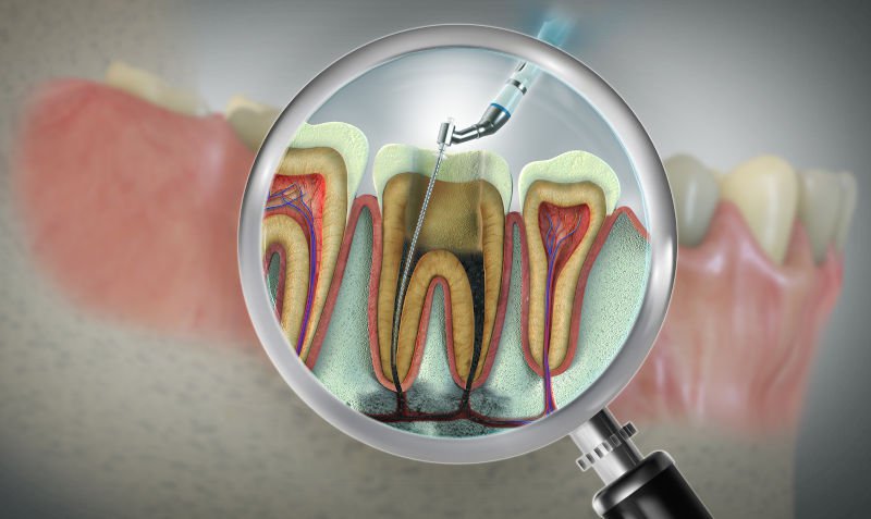
During a root canal, a dentist removes the infected or damaged pulp from inside the tooth, cleans and shapes each root canal with fine instruments, fills the space with a biocompatible material, and seals the tooth before placing a protective crown. The procedure typically takes one to two appointments of 60 to 90 minutes each, is performed under local anesthetic so you feel pressure but not pain, and saves the natural tooth from extraction. Most patients are surprised by how routine and manageable the experience is compared to what they expected.
Key Takeaways
- A root canal involves removing infected pulp, cleaning and shaping the canals, filling them with gutta-percha, and sealing the tooth with a crown. The whole process usually spans two appointments.
- Local anesthetic is used throughout. The procedure itself is not painful; patients feel pressure and vibration, not sharp pain.
- Different teeth have different numbers of root canals: front teeth typically have one, while upper molars can have three or four, making them more complex to treat.
- A root canal saves your natural tooth, preserves the jawbone, and is nearly always preferable to extraction from both a health and cost standpoint.
- Patients in Maple and Vaughan can book a root canal treatment consultation at Mapleridge Dentistry for same-day assessment when pain or swelling is present.
What Is a Root Canal and Why Is It Needed?
Before looking at how a root canal works step by step, it helps to understand the anatomy that makes the procedure necessary and the conditions that require it.
The Anatomy of a Tooth and Its Root Canals
Every tooth has three main layers: the hard outer enamel, a layer of dentine beneath it, and the pulp at the centre. The pulp is a soft tissue containing nerves, blood vessels, and connective tissue that runs from the crown of the tooth down through narrow channels in each root. These channels are the root canals. They connect to the surrounding bone and tissue at small openings near the root tips, called apices. In healthy teeth, the pulp is protected inside a sealed chamber. When bacteria reach the pulp through deep decay, a crack, or a failed restoration, the tissue becomes inflamed and eventually infected, and the pain signals sent by the pulp's nerve fibres can become severe.
What Causes a Root Canal Infection?
The most common reasons pulp becomes infected or necrotic (dead) include:
- Deep tooth decay that penetrates through the enamel and dentine and reaches the pulp chamber
- A cracked or fractured tooth that allows bacteria to enter, even without visible decay
- A large filling or repeated dental procedures on the same tooth that have gradually irritated the pulp over time
- Trauma to the tooth, such as a blow to the face, can damage the pulp's blood supply and cause the tissue to die slowly
- Severe gum disease that has allowed bacteria to travel down the root surface to the apex
If you are experiencing persistent tooth pain, sensitivity to heat or cold that lingers after the source is removed, swelling in the gum near a tooth, or a tooth that has darkened in colour, these are warning signs that the pulp may be affected and that emergency dental assessment in Maple is advisable before the infection spreads.
How Does a Root Canal Work? The Procedure Explained
The following is a detailed walkthrough of how root canal treatment in Vaughan proceeds from diagnosis to final restoration. Understanding each stage helps patients feel prepared and reduces the anxiety that comes from the unknown.
Step 1: Diagnosis and Pre-Treatment Imaging
The dentist reviews periapical X-rays or, in more complex cases, a cone beam CT scan to map the root canal system of the affected tooth. This imaging shows how many canals are present, the length and curvature of each root, and the extent of any infection or bone changes at the root tip. If a significant infection is confirmed, a short course of antibiotics may be prescribed prior to treatment to reduce the bacterial load and improve the effectiveness of the local anesthetic.
Step 2: Anesthesia and Isolation
A local anesthetic is injected into the gum and bone surrounding the tooth. Within two to three minutes, the entire area is numb. A thin sheet of latex or nitrile rubber called a rubber dam is then stretched over the tooth and clipped in place. The dam isolates the tooth from saliva, keeps the working field dry, and prevents any irrigating solutions or small instruments from entering the throat. For patients with significant dental anxiety, sedation dentistry in Maple is available as an adjunct to local anesthesia, allowing the procedure to be completed in a more relaxed state.
Step 3: Accessing and Cleaning the Root Canals
The dentist uses a dental drill to create a small access opening in the biting surface of a back tooth or through the back of a front tooth. Through this opening, the pulp chamber is entered, and each canal is located using small hand or rotary instruments. The infected and necrotic pulp tissue is removed using a series of progressively larger nickel-titanium files. Each file is worked to a precisely measured length, determined by an electronic apex locator, to ensure cleaning reaches the very tip of the root without perforating it. Between instruments, the canals are flushed with sodium hypochlorite, which dissolves organic tissue and kills bacteria, and with EDTA, which removes the calcium-containing smear layer from the canal walls, allowing the filling material to seal properly.
Step 4: Shaping, Disinfecting, and Filling
Once the canals are clean and shaped to a smooth, tapered form, they are dried completely with paper points. The dentist then fills each canal from the root tip upward with gutta-percha, a rubber-like thermoplastic material that has been the standard root canal filling for over a century due to its biocompatibility, dimensional stability, and ability to seal the canal space. A sealer cement is applied alongside the gutta-percha to fill any voids. The gutta-percha is compacted to ensure a dense, three-dimensional fill that prevents bacteria from re-entering the canal system.
Step 5: Sealing and Crown Placement
A temporary filling is placed to close the access cavity at the end of the first appointment. At the follow-up visit, the temporary material is removed, and the tooth is prepared for a permanent restoration. In the vast majority of cases, a dental crown in Vaughan is placed over the treated tooth. Back teeth that bear chewing force are particularly vulnerable to fracture after root canal treatment because the procedure removes internal structure, and a crown restores both strength and function. Front teeth with minimal structural loss may be restored with a composite filling and, in some cases, a post, but the treating dentist will advise on the most appropriate option based on how much natural tooth structure remains.
"One of the most common things patients tell me after a root canal is that they wish they had come in sooner. The procedure itself, once the anesthetic is working, is genuinely low-discomfort. What patients feel during treatment is mostly pressure and instrument movement. The pain that brings them in, from the infected pulp pressing against surrounding tissue, is almost always worse than anything they experience in the chair." Dr. James Tanzil, General Dentist, Mapleridge Dentistry
What Does a Root Canal Feel Like? Pain, Comfort, and Common Concerns
Anxiety about root canal treatment is almost universal, yet consistently contradicted by patient experience. Understanding what the procedure actually feels like helps separate the reality from the reputation.
Does a Root Canal Hurt?
A root canal performed under adequate local anesthesia is not painful. The needle used to deliver the anesthetic produces a brief sting, and some patients notice a dull ache when the instrument approaches the root tip if the infection has lowered the tissue pH and reduced anesthetic effectiveness. In these cases, the dentist tops up the anesthetic before continuing. After the appointment, mild jaw soreness and some tenderness when biting are normal for two to four days and can be managed comfortably with over-the-counter ibuprofen or acetaminophen. Once the pulp is removed, the tooth no longer has a nerve to transmit pain, so the long-term result is a tooth that is permanently pain-free.
How Long Does a Root Canal Take?
A straightforward root canal on a front tooth with a single canal typically takes 45 to 60 minutes. Molars with three or four canals take 60 to 90 minutes and sometimes require a second cleaning appointment before the final fill if the infection was severe. Most patients complete the full treatment, including the permanent crown, across two appointments. Same-day treatment is occasionally possible for single-canal teeth where no active infection is present.
Teeth Root Canals: How Many Canals Does Each Tooth Have?
The number of root canals in a tooth varies by tooth type and, to some degree, by individual anatomy. This is clinically significant because missing a canal during treatment leaves infected tissue in place and is one of the most common reasons root canal treatment fails. The table below outlines the typical canal count for each tooth type in the mouth.
| Tooth Type | Typical Number of Root Canals | Clinical Note |
|---|---|---|
| Upper front teeth (central and lateral incisors) | 1 | Single straight canal; relatively straightforward procedure |
| Upper canine | 1 | Longest root in the mouth; the canal can be narrow, but usually singular |
| Upper premolars (bicuspids) | 1 to 2 | The first upper premolar frequently has two canals; the second may have one |
| Upper molars | 3 to 4 | Three roots are common; the mesiobuccal root often has two canals, making upper molars the most complex to treat |
| Lower front teeth (incisors) | 1 to 2 | Smallest teeth; may have two canals that merge near the root tip |
| Lower canine | 1 | Similar to the upper canine, generally one canal |
| Lower premolars | 1 to 2 | Variable anatomy; can have one, two, or rarely three canals |
| Lower molars | 2 to 3 | Two roots, each typically containing one to two canals; the total is usually three |
Upper molars are statistically the most complex teeth for root canal treatment because the mesiobuccal root frequently has two canals that are close together and can be missed on standard X-rays. Cone beam CT imaging is increasingly used for these teeth to confirm the number of canals before treatment begins. Anatomical variation means that the counts above are typical ranges, not guarantees, and thorough imaging before and during treatment is always part of a well-executed root canal.
Signs You May Need a Root Canal
Not every toothache requires a root canal, but certain symptom patterns reliably indicate that the pulp is compromised and treatment is likely needed. The most common warning signs include:
- Severe, throbbing tooth pain that is continuous or that wakes you from sleep
- Sensitivity to hot or cold temperatures that persists for more than 30 seconds after the stimulus is removed
- Pain when biting down or applying pressure to the tooth
- Swelling or a raised bump on the gum near the affected tooth, sometimes with a small draining sinus
- Tooth discolouration, particularly a greyish or darkening shade compared to adjacent teeth
- A cracked or chipped tooth that has been accompanied by new or worsening pain
A tooth that has become abscessed and is causing significant swelling or systemic symptoms, such as fever, should be assessed urgently. Regular dental visits allow dentists to catch deep decay or cracks before the pulp is affected, which is why maintaining routine checkups through consistent preventive dental care in Vaughan is one of the most effective ways to avoid root canal treatment altogether.
Root Canal vs Tooth Extraction: Which Is Better?
When a tooth is severely infected, patients are sometimes offered a choice between root canal treatment and extraction. In the vast majority of clinical situations, saving the natural tooth through root canal treatment produces better long-term outcomes.
A natural tooth transmits chewing forces into the jawbone, which stimulates bone maintenance. When a tooth is extracted and not replaced, the surrounding bone begins to resorb within months. The adjacent and opposing teeth gradually shift toward the gap, altering the bite and increasing the risk of further dental problems. Replacement options following extraction include dental implants, a fixed bridge, or a partial denture. A dental implant in Vaughan is the most functionally similar replacement to a natural tooth, but it requires a surgical procedure, a healing period of several months, and a significantly higher total cost than root canal treatment and a crown.
Extraction is the more appropriate choice when the tooth is unsalvageable due to severe structural loss, when the surrounding bone has been extensively destroyed by infection, or when the patient's medical history makes a root canal impractical. In these situations, the treating dentist will discuss dental implant options in Vaughan as part of a replacement plan. For most patients, however, root canal treatment is the more conservative, cost-effective, and clinically sound option.
Recovery After a Root Canal
Recovery from root canal treatment is typically straightforward. The following guidelines help the treated tooth heal correctly and avoid complications.
- Avoid chewing on the treated tooth until the permanent crown is placed, as an uncrowned tooth with an access cavity is more vulnerable to fracture
- Take over-the-counter anti-inflammatory pain relief such as ibuprofen for the first two to three days if soreness is present, as directed on the packaging
- Maintain normal oral hygiene, including brushing and flossing; the treated tooth can be brushed as usual
- Avoid very hard or crunchy foods on the side of the treated tooth until the crown is fitted
- Contact the dental office if swelling increases after the first 48 hours, if you develop a fever, or if the temporary filling feels loose or falls out
Once the permanent crown is placed, the treated tooth functions the same as any other tooth in the mouth. A root-canal-treated tooth can last for decades with normal oral hygiene and regular dental check-ups. There is no maintenance specific to a root canal-treated tooth beyond what is recommended for all teeth.
Book Your Root Canal Consultation at Mapleridge Dentistry in Maple
If you are experiencing tooth pain, sensitivity, swelling, or any of the signs described in this article, our team at Mapleridge Dentistry is ready to help. We offer same-day emergency assessment for patients with active dental pain, and our dentists near me in Maple and Vaughan are experienced in root canal treatment across all tooth types, including complex multi-canal molars.
Early treatment nearly always means a more straightforward procedure, faster recovery, and a better long-term outcome for the tooth. Do not wait for the pain to escalate. Book your consultation today or contact our Maple office to speak with our team.
For patients who are up to date with their preventive dental care in Vaughan, mention any tooth sensitivity or discomfort at your next scheduled exam so it can be assessed before a root canal becomes necessary.
Frequently Asked Questions
What happens in a root canal?
During a root canal, the dentist removes infected or dead pulp tissue from inside the tooth, cleans and disinfects each root canal with instruments and irrigating solution, fills the empty canals with gutta-percha, and seals the tooth. A crown is then placed to restore the tooth's shape and strength. The full process is performed under local anesthesia and typically requires two appointments. More information about root canal treatment in Maple is available on our service page.
How does a root canal work at the biological level?
Root canal treatment works by eliminating the source of infection inside the tooth. Bacteria that have colonized the pulp space produce toxins that inflame the surrounding bone and tissue. By mechanically removing the infected pulp, chemically disinfecting the canal walls, and sealing the space to prevent bacterial re-entry, the procedure allows the surrounding tissues to heal. The bone at the root tip that was damaged by the infection typically regenerates over the following months, as shown on follow-up X-rays.
What happens during a root canal that makes patients nervous?
Most anxiety about root canal treatment is driven by the unfamiliar sounds and sensations of the procedure rather than actual pain. The drill used to open the tooth, the vibration from the files, and the time the mouth needs to stay open are the most commonly reported sources of discomfort. None of these involves pain when the anesthetic is working. Patients who experience significant dental anxiety can discuss sedation options with the dentist before the appointment so that appropriate support is in place.
How do they do a root canal on a back tooth?
The approach for a back tooth is the same as for any other tooth, but back teeth present additional complexity because they have more roots and more canals, and the access opening is made in the biting surface rather than through the back of the tooth. Molars, in particular, may have three or four canals, each of which needs to be located, cleaned, and filled individually. The appointment is longer than for a front tooth, typically 75 to 90 minutes, and more advanced imaging may be used before treatment to confirm the canal anatomy.
How long does a root canal take from start to finish?
A root canal on a single-canal front tooth can be completed in one 45 to 60-minute appointment. Multi-canal back teeth require 60 to 90 minutes per cleaning appointment, and sometimes two cleaning visits if the infection was significant, before the fill and temporary seal are placed. The permanent crown is fitted at a separate visit, usually two to three weeks after the root canal is completed. Total treatment time from first appointment to final crown placement is typically three to five weeks.
